By Dr. Dina Elsalamony MD, MScPH
The feet are the most distal part of the human leg, they function to balance our posture, transmit our weight to the ground, and help us move accurately — our heels are the first point of contact between the ground and our bodies. The heel (or the calcaneus bone) is cushioned and supported by a layer of fat known as the calcaneal fat pad, which is normally about 1-2 cm in thickness with the average healthy calcaneal fat pad measuring around 18 mm thick.
What is the heel fat pad?
The heel fat pad is made up of fatty tissues that are enclosed by ligamentous chambers. During the human gait cycle of walking, standing, jumping, and running, our heel pads act as cushions protecting our heel bones, nerves and blood vessels from damage by absorbing the shock of the impact of our feet on the ground. Our heel fat pad also serves as a mechanical anchor that helps to distribute our weight appropriately without putting too much pressure on the underlying tissues.
Our feet endure a lifetime of excessive use, it is thought that the human feet travel over 100,000 miles in about 75 years. The heel can absorb 110% of the body’s weight during walking and 200% of the body’s weight during running; this excess mileage and chronic increase in pressure strike and load forces could result in thinning of the heel fat pad, leading to a common complain of heel pain.
In addition to the natural degenerative process, a number of other factors could affect this layer of fat on our heels leading to its thinning or displacement, compromising the protection of the underlying heel bones which could result in heel pain, and sometimes disability. This condition is referred to as heel pad syndrome, in this article we will discuss everything you need to know about this condition and the possible treatments.
What is heel fat pad syndrome?
Heel fat pad syndrome is a condition that happens as a result of changes in the elasticity and/or the thickness of the heel fat pad, this is often caused by wear and tear over time of the fatty tissues that make up the heel pads on our feet causing pain that could impact our daily routine and interfere with our regular activities.
The atrophy of the heel pad is considered to be the second leading cause (14.8%) of plantar heel pain (1) after plantar fasciitis.
Risk factors and causes of heel fat pad syndrome:
Various factors can play a role in developing heel pad syndrome, and in a lot of cases, this condition could be attributed to a combination of a number of factors.
These factors include:
- Aging: As we age, many of our body systems go through a degenerative process in which they become less effective, this applies to everything including our soft tissues. The process of aging affects the thickness and elasticity of our feet heel fat pads. As we get older and with use, more commonly after the age of 40 years old, the fatty tissues cushioning our heel start to break down, with loss of collagen, water, and elastic tissues, reducing the shock absorbency and leaving us more susceptible to pain and heel bone bruising.
- Trauma: Injury of the heel fat pad can lead to heel pad syndrome; the injury could be a result of trauma to the heel while playing high impact sports/ activity or as a result of a direct blow to the heel such as in the case of car accidents.
- Obesity: The higher the body weight the more stress it puts on the heel fat pads, causing faster loss of elasticity and the shock absorber properties of the fat pad. (2)
- Issues with structure and gait: conditions, injuries, or deformities to the feet, ankle, knee, or hips can alter the walking pattern of the individual leading to uneven weight distribution across the heel, which eventually results in aggravated breaking down of the heel fat pad.
- Improper footwear: wearing inappropriate shoes that don’t provide enough support, particularly when performing weight-bearing activities or high impact exercises can exacerbate the loss of the heel pads. Prolonged wearing of high heels or walking barefoot can also contribute to the development of this condition.
- Hard surfaces: walking or exercising on hard surfaces increases the shock that the heel receives on impact, increasing the possibility of thinning heel fat pads.
- Repetitive activities: any activities that include extended and/or high impact on the heel such as running, jumping, gymnastics, and prolonged standing or walking, can cause chronic overload on the fatty tissues of the heel pad leading to inflammation and a higher risk of developing heel pad syndrome.
- Corticosteroid injections: atrophy of the heel fat pad can sometimes be a side effect of corticosteroid injection in the feet as part of the treatment of pain and inflammation from other feet conditions. (3)
- Plantar fasciitis: The degeneration of the plantar fascia and its impaired ability to absorb the shock and support the feet, can result in putting more load on the heel and causing heel pad syndrome.
- High arch (cavus foot): People who have high arched feet are more likely to develop fat pad syndrome, particularly if they are runners, as the fat pad becomes displaced with use.
- Genetics or family history: are factors that could be strongly associated with excessive fat pad loss or degenerative diseases.
- Other health conditions: such, as rheumatoid arthritis and lupus can affect the connective tissue in the feet. Diabetes mellitus has also been associated with higher incidences of heel pad syndrome due to adipose tissue atrophy and degeneration of the collagen in this area, (4) additionally, in the case of those who suffer from diabetic peripheral neuropathy (damage of the peripheral nerves supplying the legs and feet), they tend to lose some of the sensations in the feet leading to inability to feel pain, so they continue to put too much pressure on the heel, which in return worsens the atrophy of the heel fat pads.
What are the symptoms of heel fat pad syndrome?
If you suffer from heel fat pad syndrome, you will likely experience the following symptoms:
- Deep dull aching pain in the heel, typically in the middle of the calcaneus bone which would be around the center of the heel.
- The pain experienced is often felt as if there was a bruise in the heel pad when pressing on the heel while walking, running or standing for a long time.
- The pain could be recreated by pressing at the center of the heel.
- This pain increases in severity by prolonged walking or standing, particularly when barefoot, walking on hard surfaces, or while performing any high-impact exercise.
- Less common symptoms could include tingling, cold or burning sensation which tend to be a result of a neurological cause as nerves that are located in the superficial regions of the heel can be easily compressed after atrophy of the heel fat pad. (1)
- Mild cases of heel pad syndrome might be asymptomatic, or felt occasionally when aggravated by pressing on it directly or came on contact with hard surfaces such as hardwood floors, concrete or ceramic tiles.
How is heel pad syndrome diagnosed?
If you think that you might have heel fat pad syndrome, it is important to see your physician to get an accurate diagnosis and proper management for this condition.
Your physician will first ask you about your medical history if you have any other medical conditions that could cause heel pain or contribute to the loss of fat in your heels. He or she will ask you to describe in detail the type of pain that you get, when did you first notice the pain? What time of the day do you get it most? Does it get better with rest?
Your doctor will then move to perform a physical examination, to properly examine your feet for any change of skin appearance or for structural deformity, in addition to attempting to replicate the pain by pushing with their fingers on the bottom of the feet until they can localize the exact area of the pain if possible. On some occasions, the bones could be felt through the skin.
Another examination could be for the evaluation of the heel pad elasticity, by measuring the change in the thickness of the heel pad when your feet are supporting your weight while standing versus when they are not while you are sitting or lying down. A sign of low elasticity could be detected if your heel fat pad is stiff and does not properly compress when supporting your weight.
Additionally, your physician may require X-ray to exclude other conditions that have similar presentation such as plantar fasciitis or stress fracture of the heel bone.
In most cases of heel fat pad syndrome, clinical diagnosis using the above techniques is the standard, however, when clinical examination is not conclusive, ultrasound or MRI could be reserved for these situations as they are also great diagnostic tools for this condition.
What is the treatment of heel fat pad syndrome?
The management and treatment of this condition could be categorized into two approaches:
Conservative treatment for fat pad synrome
In this approach, the goal is to manage the symptoms by decreasing the pain and inflammation and improve the quality of the individual’s life. Conservative treatment does not include replacing the lost fat tissues in the heel pad, instead, it strives to preserve and protect the number of fatty tissues left in the heel pad, prevent further damage and decrease inflammation which in return allows the fatty tissues to possibly recover through the natural healing process.
These treatments include:
- Rest: it is important to rest your feet when you experience such pain, try to avoid high-impact sports, carrying heavy loads, and minimize the time you stay standing or walking.
- Icing: Icing your feet can help reducing the inflammation and the pain caused by heel pad syndrome. Try to use an ice pack for 20 minutes after every activity that may cause pain. Heel That Pain offers Ice Therapy Slippers that are extremely convenient and easy to use, providing a practical and comfortable solution to heel icing. The Ice Therapy Slippers ensure total covering of the bottom of the feet easing the inflammation and relieving heel pain.
- Manual therapy and exercises: Physical therapy that includes rehabilitation exercises for the joints and muscles of the feet, ankles, upper and lower legs. Heel soft tissue manual massage therapy has also been shown to be effective in relieving some of the pain associated with heel pad syndrome.
- Therapeutic low-dye taping: This is a well-established technique in the treatment of heel pain, more commonly used for plantar fasciitis than fat pad atrophy, however multiple studies (5,6,7) have reported great results in using this technique in treating fat pad syndrome. The mechanism of taping relies on producing an anti-pronation effect reducing the heel pain, dispersing pressure under the plantar area, (8) and providing a cushioning effect on the heel by helping to increase the thickness of the heel fat pad. This technique is useful, particularly indoors where it is less convenient to wear orthotics and heel cushions. Taping has a variety of modified versions, and one of the most commonly used modifications that is proven more effective in treating heel pad atrophy is adding figure-8 taping around the heel area. (9,10)
- Medication: Over the counter pain relief and anti-inflammatory medications such as Voltaren and Ibuprofen, can often help in reducing the heel pain.
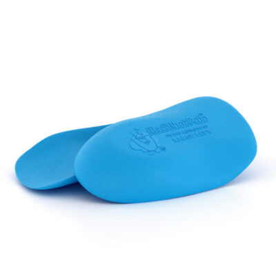
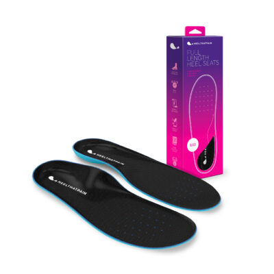
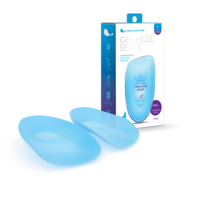
- Orthotics and heel cups: One of the most effective conservative techniques in treating heel fat pad syndrome is the use of shoe inserts and cups. (11) The idea is to offload the heel area by providing sufficient cushioning and support to reinforce the thinning and stiff heel fat pad, leading to better distribution of weight and reduction of the pressure on the heel bones and surrounding structures. The Heel That Pain Heel Seats utilizes the unique and revolutionary technology of fascia bar that absorbs the impact when standing, walking or running, providing excellent support for the heel. Heel seats use a dense cushion of soft Keraton that repels moisture, sweat and bacteria and prevents sliding inside the shoes when moving. Additionally, Heel Seats apply gentle therapeutic acupressure to the heel bone, which encourages the natural human body healing process. This is a great affordable and effective option that could be purchased conveniently online without the need of prescription or custom sizing as it it’s available in various sizes, in addition to a gel version if your feet are too sensitive for the standard firm option.
Advanced treatments for fat pad syndrome:
If conservative treatments have failed to improve the heel fat pad condition and no increase in the heel fat pad thickness have been observed, or in the case of severe atrophy that greatly compromises the underlying heel bone structure and/or superficial nerves, more advanced treatment could be suggested by your physician with the aim of replacing the thinning heel fat pad to provide faster more noticeable results and prevent any more damage. The different fat replacement techniques have various degrees of success and could all fall under the term of heel pad augmentation, which could be broken down into the following three categories:
- Injectables: There are various injectable materials that are used to thicken the atrophied or displaced fat pad in the heel, these materials are referred to as dermal fillers. Dermal fillers are made different kinds of either natural or synthetic materials that are injected into the skin to increase volume of a specific area on the body. The use of dermal fillers containing materials such as poly-L-lactic acid and hyaluronic acid, for treating of fat pad atrophy is one of the techniques that are becoming increasingly popular despite being considered as off-label use by physicians (meaning derma fillers use is indicated and approved for treatment of wrinkles but they are being used by physicians in this case for treatment of fat pad syndrome which is not the original indication for their use), this is because they provide easy and quick, short to intermediate-term solution to this condition. They usually have no to minimal side effects, which could include minor bruising, tenderness and localised inflammation. The recovery after the procedure is quick as the person can start walking on their feet immediately after, providing initially wearing footwear or shoe inserts and avoiding high-impact activities. The results usually can last from 6 months to 2 years depending on the material used in the filler, the age and weight of the person, their life style and activity and the degree of atrophy that they were presented with initially. On the other hand, the most controversial injectable material is silicone as it has the tendency to migrate after a while from one place in the body to another in some cases which could cause negative side effects. Additionally, silicone can also induce a foreign body reaction that could range from mild to severe. (12)
- Autolipotransplantation: also known as fat grafting or autologous fat transplantation is another yet more advanced augmentation technique that involves taking fatty tissues from one part of the body and placing it into another, in this case the heel. The physician would normally take the fat from one area that has a high value of fat such as the thighs or the abdomen and transfer it to the graft location at the bottom of the foot, only a small amount of fat (few cubic centimetres) is needed for this procedure. Local anesthesia is used at both the harvest and graft sites to avoid pain and bruising during the procedure. Autograft procedures to fill the heel fat pad are usually quick and don’t require hospitalization as they can be performed at the doctor’s office or the outpatient clinic, and the person would be discharged in the same day. The recovery time is slightly longer with this option as the feet would be bandaged for 24hrs after the procedure, and for 3 weeks after great care should be taken when it comes to the footwear and the activities performed. The success rate for treating heel fat pad syndrome using this technique is 98%, (13) with results lasting for much longer period of time than the injectable derma fillers, although it is advised to continue wearing sensible cushioned shoes and minimise high and chronic weight-bearing activities to ensure long-lasting results.
- Allografting: Which means augmentation of the heel fat pad using fat obtained from another person, this fat graft does not contain living cells to ensure that body of the receiver would not reject the it. This procedure is more complicated and require a surgical setting to be performed, with long recovery time ranging from 6 to 8 weeks since the graft take time to incorporate with the person’s own tissue, but once incorporated, the results are remarkable with significant relief of pain and other symptoms for about 5 years or more.
Treatment of deformity:
If the cause of heel fat pad syndrome is an underlying deformity that puts too much weight and pressure on the heel, it is important to address this deformity in addition to locally treating the heel fat pad, that is to ensure low chance of recurrence. For example, people with diabetes who developed claw toe deformity, or those with ankle alignment issues, will need to treat the deformity as well as the heel fat pad, the treatment could be conservatively or surgically if required.
How to prevent heel fat pad syndrome?
As mentioned above, there are many factors that could contribute to developing this condition, of which some cannot be avoided such as age or genetics, however plenty of lifestyle and occupational causes could be negated if the appropriate measures are being taken. These include:
- Ensuring proper footwear that is densely cushioned and provide enough support to the bottom of the feet at all times.
- Wearing athletic shoes when performing any high-impact activity.
- Limiting the intensity and duration of high-impact and weight-bearing activity, such as running or prolonged standing, to ensure the heel fat pad’s ability to recover after the activity.
- Try to avoid wearing high heels, particularly pointy heels, as high heels can poorly distribute the weight of your body, putting too much pressure on one area of your feet.
- Avoid walking barefoot, especially on hard or uneven surfaces.
- Inspect your athletic shoes regularly and change them when you observe a noticeable reduction in the cushioning.
- Maintain a healthy weight.
Plantar Fasciitis vs. heel fat pad syndrome:
Heel fat pad syndrome is commonly misdiagnosed at plantar fasciitis, while both can be a source of heel pain, the risk factors, symptoms, and treatment vary for each condition.
The location of the plantar fascia plays a part in heel fat pad syndrome being mistaken for plantar fasciitis, as one end of the plantar fascia is attached to the inside part of the heel and there is an overlap where the part is covered by the heel fat pad, the rest of the plantar fascia travels on the bottom of the feet forming the medial longitudinal arch. This arch provides dynamic shock absorption and static support of the feet during movement, while the heel fat pad is responsible more for the absorption of the initial impact when the heel comes in to contact with the ground. When heel pad atrophy occurs, destruction of the fibrous membrane in the heel pad starts to happen, in addition to the loss of moister and soft tissue elasticity reduction leading to impaired shock absorption mechanism. (14)
Plantar fasciitis is a condition that happens due to chronic degeneration of the plantar fascia fibers, affecting the blood flow to the plantar fascia leading to its inability to naturally repair and heal itself. This degenerative nature has been attributed to repeated microtrauma to the plantar fascia that occurs as a result of the sudden increase of intensity or length of weight-bearing exercises, fast weight gain, prolonged standing, or due to structural deformities of the joints, bones, or muscles of the individual leading to unloading too much weight and pressure on the plantar fascia causing chronic microtraumas. (15)
The symptoms of plantar fasciitis vary from those of fat pad syndrome as in the case of plantar fasciitis the pain is usually described as dull aching or throbbing pain located at the inner front portion of the heel which extends to the foot arch, where this part commonly becomes tender or swollen. The plantar fascia would sometimes also feel tight particularly during stretching, additionally, one of the most characteristic signs of plantar fasciitis is that the pain usually happens when the person takes their first step after a long period of resting, such as the first step in the morning getting out of bed; the pain usually decreases after walking on their feet for few minutes, but often comes back if the person continued walking for a long period. On the other hand, the pain of fat pad syndrome the pain is described more as deep dull aching pain that feels like a bruise in the heel bone, the pain is at the center of the heel and happens usually while walking or standing for a long time, or when walking barefoot and is aggravated by walking on hard surfaces. It is more common to have pain in both feet in the case of fat pad syndrome than in plantar fasciitis where it is more likely to occur in one foot, (7) the pain also happens more often during the night than in the case of plantar fasciitis when it is experienced first thing in the morning.
On some occasions, the person could suffer from both plantar fasciitis and heel fat pad syndrome at the same time, which makes it even more challenging to clinically diagnose, therefore when there is suspension and inability to confirm the diagnosis of fat pad syndrome or/and plantar fasciitis, the use of ultrasound and MRI is recommended to confirm the condition and prescribe the appropriate treatment accordingly.
Final word of advice:
If you experience heel pain, it is really important to consult with your physician to determine the possible cause and address any underlying issues. The earlier the diagnosis and management of any heel condition the better the prognosis and less damage that could occur to any of the feet structures as a consequence. Always ensure wearing appropriate shoes that offer support and cushioning for your heel and the bottom of your feet, and take it easy when it comes to high-impact, weight-bearing activities that could put too much stress and pressure on the bottom of your feet and your lower limb joints.
REFERENCES:
- Yi, T. I., Lee, G. E., Seo, I. S., Huh, W. S., Yoon, T. H., & Kim, B. R. (2011). Clinical characteristics of the causes of plantar heel pain. Annals of rehabilitation medicine, 35(4), 507–513. https://doi.org/10.5535/arm.2011.35.4.507
- Aldridge T. (2004). Diagnosing heel pain in adults. American family physician, 70(2), 332–338.
- Yucel I, Ozturan KE, Demiraran Y, Degirmenci E, Kaynak G. (2010). Comparison of high-dose extracorporeal shockwave therapy and intralesional corticosteroid injection in the treatment of plantar fasciitis. J Am Podiatr Med Assoc, 100 (2):105-10. [Medline].
- Hsu CC, Tsai WC, HsiaoTY, Tseng FY, Shau YW, Wang CLet al. (2009). Diabetic effects on microchambers and macrochambers tissue propertiesin human heel pads. Clin Biomech, 24:682-686.
- Yi TI, Shim JW, Ku HK, Seo IS, Huh WS, Kim JS, et al. (2011). The effects of modified low-dye taping in the patient with heel pad atrophy. J Korean Acad Rehabil Med, 35:96–100. [Google Scholar]
- Ha HS, Hwang JH, Kim YB, Ha YM, Hwang JM. (2012). Immediate clinical and biomechanical effects of low dye taping in patients with plantar heel pain. Korean J Sports Med, 30:9–15. [Google Scholar]
- Chae, Y. H., Kim, J. S., Kang, Y., Kim, H. Y., & Yi, T. I. (2018). Clinical and Biomechanical Effects of Low-Dye Taping and Figure-8 Modification of Low-Dye Taping in Patients With Heel Pad Atrophy. Annals of rehabilitation medicine, 42(2), 222–228. https://doi.org/10.5535/arm.2018.42.2.222
- Pfeffer, G., Bacchetti, P., Deland, J., Lewis, A., Anderson, R., Davis, W., Alvarez, R., Brodsky, J., Cooper, P., Frey, C., Herrick, R., Myerson, M., Sammarco, J., Janecki, C., Ross, S., Bowman, M., & Smith, R. (1999). Comparison of custom and prefabricated orthoses in the initial treatment of proximal plantar fasciitis. Foot & ankle international, 20(4), 214–221. https://doi.org/10.1177/107110079902000402
- Radford, J. A., Burns, J., Buchbinder, R., Landorf, K. B., & Cook, C. (2006). The effect of low-Dye taping on kinematic, kinetic, and electromyographic variables: a systematic review. The Journal of orthopaedic and sports physical therapy, 36(4), 232–241. https://doi.org/10.2519/jospt.2006.36.4.232
- Newell, T., Simon, J., & Docherty, C. L. (2015). Arch-Taping Techniques for Altering Navicular Height and Plantar Pressures During Activity. Journal of athletic training, 50(8), 825–832. https://doi.org/10.4085/1062-6050-50.5.05
- Chanda, A., & McClain, S. (2019). Mechanical Modeling of Healthy and Diseased Calcaneal Fat Pad Surrogates. Biomimetics (Basel, Switzerland), 4(1), 1. https://doi.org/10.3390/biomimetics4010001
- Altmeyer, M. D., Anderson, L. L., & Wang, A. R. (2009). Silicone migration and granuloma formation. Journal of cosmetic dermatology, 8(2), 92–97. https://doi.org/10.1111/j.1473-2165.2009.00436.x
- Chairman EL. Restoration of the plantar fat pad with autolipotransplantation. J Foot Ankle Surg. 1994; 33(4):373-9.
- Turgut, A., Göktürk, E., Köse, N., Seber, S., Hazer, B., & Günal, I. (1999). The relationship of heel pad elasticity and plantar heel pain. Clinical orthopaedics and related research, (360), 191–196. https://doi.org/10.1097/00003086-199903000-00023
- Riddle DL, Pulisic M, Pidcoe P, Johnson RE. (2003). Risk factors for Plantar fasciitis: a matched case-control study. J Bone Joint Surg Am, 85-A(5):872-7. [Medline]
Fat Pad FAQ
The heel fat pad is exactly as it sounds — a pad of fatty tissues enclosed by. ligamentous chambers on the heel of your foot. It cushions your heel bones, nerves, and blood vessels throughout normal activities like walking, standing, and jumping.
Heel fat pad syndrome is a condition that occurs after long-term wear and tear of the fatty tissues of the heel. Over time, the fat pad can wear out and cause pain.
The first steps towards improving pain from fat pad atrophy are rest, icing, and orthotics. You can also try over-the-counter anti-inflammatories like ibuprofen. Advanced medical treatments for fat pad syndrome include injectables and fat grafting.
Fat pad syndrome and plantar fasciitis are often confused. Plantar fasciitis is a degenerative condition that affects the plantar fascia ligament, while fat pad syndrome involves the atrophy of the fatty tissue layer protecting the heel bone. Both conditions can cause significant heel pain.


